Post-traumatic arthritis of the knee: when injury is followed by disease
- 1 Post-traumatic knee osteoarthritis
- 2 Post-traumatic knee arthritis develops
- 3 Post-traumatic osteoarthritis of the knee
- 4 Post-traumatic osteoarthritis of the knee
- 5 Post-traumatic knee osteoarthritis
- 6 Post-traumatic knee osteoarthritis
What is it?
Arthrosis of the knee joint is the most common form of osteoarthritis in Europe, along with osteoarthritis of the hip.
Most of these are consequences of “normal” wear and tear osteoarthritis. Still, more than one-tenth of arthrosis (12 percent) develops due to joint injury and, thus post-traumatic arthrosis.
The human musculoskeletal system is constantly exposed to heavy loads.
It can be damaged over the years or by accident: tendons, ligaments, meniscus, cartilage, soft tissue, and bone can be severely affected by tears or fractures. However, such acute injuries are often followed by wear and tear and, as a result, an inflammatory process called arthritis due to the passage of pain, neglect, overexertion, and hereditary disposition. After some time, this often turns into arthrosis, which describes irreversible damage to the bone.
The knee joint is particularly and frequently affected by arthrosis after intra- and extra-articular fractures.
After a tibial plateau fracture or distal femur fracture, post-traumatic arthrosis occurs with 21% to 74% within ten years. However, capsule and ligament injuries resulting in instability, meniscus injuries, or direct cartilage damage can also trigger post-traumatic osteoarthritis. Depending on the extent of the damage, post-traumatic arthrosis can be observed in more than 80% of patients in the medium term.
How it develops?
Post-traumatic arthritis (PTA) is associated with significant physical impairment and loss of function. The development of post-traumatic arthritis (PTA) often occurs after a joint fracture. However, the complexities of the body involved in its development and progression after a joint injury are still largely unknown. Any joint injury throughout life can eventually lead to post-traumatic osteoarthritis (PTOA). Bone marrow edema is often a sign that should not be overlooked!
Common causes are:
- sports injury
- falls
- motor vehicle accidents
- previous joint surgery
Injury damages cartilage or bone and can change the mechanics of the joint. These changes, in turn, cause faster wear and tear of the joint, especially if the joint is subjected to additional stresses over time. For this reason, post-traumatic osteoarthritis is more common in younger patients than typical osteoarthritis.
Accelerated degeneration of articular cartilage resulting in post-traumatic osteoarthritis
Each step exposes the knee joint to a very high mechanical load. Therefore, after injuries, anatomical reconstruction of the joint surface and restoration of an anatomical axis of the leg is critical to avoiding post-traumatic arthritis of the knee joint.
If the remaining joint step is less than 2 mm and the axis deviation is less than 8°, at least in the mid-term follow-up, arthritis rates will increase by only 6%. In addition, restoring anatomical axial relationships is a decisive factor regarding subsequent endoprosthetic treatment.
However, it is not only mechanical factors that play a role in post-traumatic osteoarthritis. Although not all factors of post-traumatic osteoarthritis are entirely understood, the combination of mechanical damage and release of pro-inflammatory cytokines (particularly interleukin-1 and tumor necrosis factor α) appears to be a major cause of the rapid development of osteoarthritis.
It is known that the modified spectrum of cytokines leads to accelerated degeneration of articular cartilage and also increases the synovial inflammatory reaction.
Because of this accelerated degeneration, the time when endoprosthetic treatment is needed, usually around the age of 62, is about ten years earlier than in the case of primary osteoarthritis.
Post-traumatic arthrosis from fractures without joint involvement
For example, fractures of the diaphysis of a long bone that subsequently heals in a less-than-ideal position can result in axial misalignment of neighboring joints. However, any randomly produced gross misalignment results in a load on at least one joint that does not fit the body and does not match the normal shape, which then wears out (as in primary arthrosis) faster than usual and evolves into very painful post-traumatic arthrosis.
Post-traumatic arthrosis from fractures involving joints
If a trauma (such as a fracture) crosses a joint surface, that injury often heals, despite all efforts, with the formation of a step in the joint. This means that damage to the cartilage is inevitable in the near future: if the cartilage is worn away, the joint bone itself wears away. Osteoarthritis develops quickly because of the trauma, which is immediately very painful and much faster than usual. The bone rubs against the bone without protection until the joint bone is completely worn out in places. In a kind of “pitting,” in the final stage, an ever-increasing hole forms in the bare joint surface, which fills with fluid.
Subchondral cyst – synonym – such arthrosis caused by severe damage to the joint bone up to the medullary cavity, so the former name already suggests a traumatic cause. At the same time, the latter also indicates normal joint wear and tear.
Knee ligament injuries
Ligament injuries often result in poor guidance in the capsular ligament apparatus of the joint. Under load, the joint surfaces may tilt against each other, the cartilage layer is overloaded, and wear and tear leads to arthrosis.
The symptoms
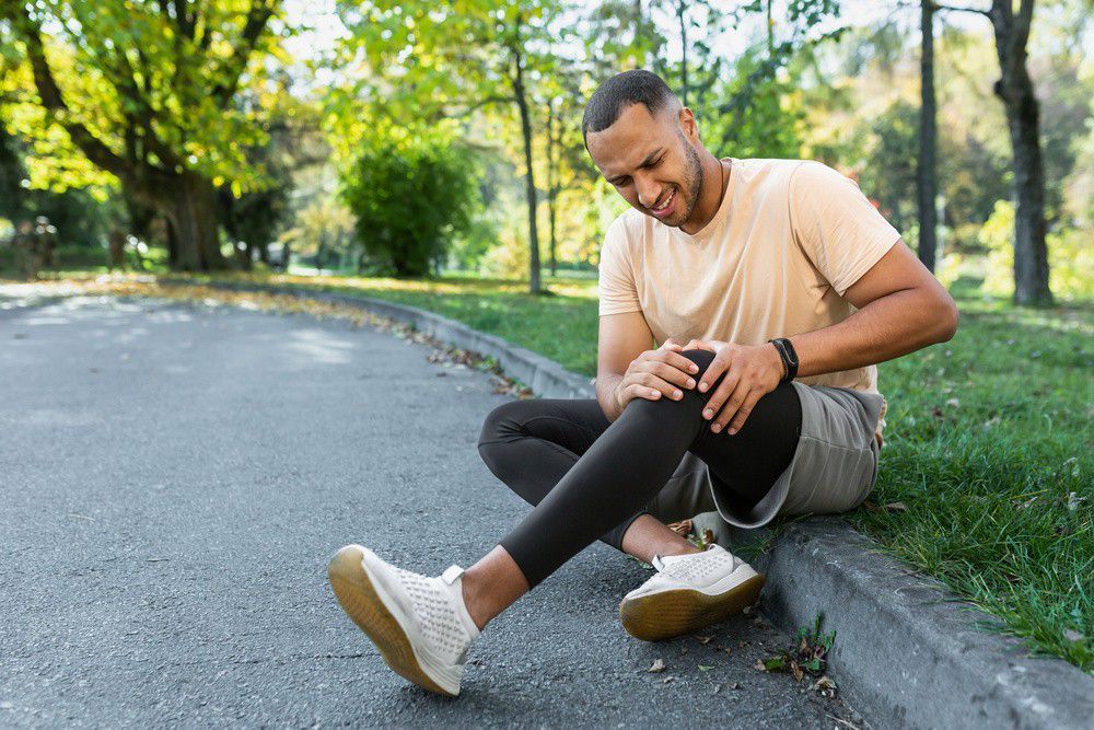
Pain and swelling in a previously damaged joint may be a symptom of post-traumatic osteoarthritis. Even injuries that are treated quickly can flare up years later and cause more rapid wear and tear on a joint. If pain occurs, it is always a good idea to consult a medical professional who will evaluate the symptoms in a detailed history.
Waiting and delaying symptoms, such as through painkillers not prescribed by a physician, is strongly discouraged!
Joint inflammation can cause permanent damage that can only be repaired surgically.
Common signs and symptoms are:
- pain or helplessness in the joint
- swelling
- joint stiffness or instability
- cysts or other abnormalities.
If a lower tolerance for knee loading is present even and only in certain positions, the physician should be consulted to be on the safe side.
The diagnosis
Clinical examination is of particular importance. In particular, an inspection of preexisting scars and soft tissue status is critical to plan follow-up care: it requires differential stability testing and range of motion.
Inclusion of history, but particularly of previous operations and their access routes with any foreign materials that may still be present, is mandatory. Radiologic diagnosis includes standard images of the knee joint and imaging of the whole leg position to analyze bony deformities and axis deviations accurately. In individual cases, prolonged exposure may also be useful to objectify ligamentous instability. CT diagnostics are recommended, especially if fracture consolidation is unclear or for more precise imaging of bony defects. For significant bone defects, CT diagnostics are especially advantageous to plan options.
In the case of complex axial misalignments and especially rotation, computed tomography can be helpful for measurement (3D planning).
The therapy
Physical therapy exercises are also recommended to strengthen the muscles around the knee joint.
In overt post-traumatic osteoarthritis caused by a previous injury, a replacement (of a part) of the joint is often the last resort to restore joint health fully.
If post-traumatic arthrosis has occurred, the natural axes of the joint can be restored, if necessary, by corrective osteotomy.
To restore an unstable ligamentous apparatus, there are various plastic surgeries where, for example, tendons are removed from another part of the body and stitched in place of the damaged ligament. For traumatic joint cartilage damage, joint cartilage surgeries often lead only to short-term improvement of symptoms.
Because each patient is very different in their daily life, needs, and clinical picture, the ideal path to a pain-free quality of life must be found together with the surgeon.
Post-traumatic osteoarthritis of the knee: surgical therapy
Treating patients with post-traumatic osteoarthritis is significantly more time-consuming, complex, and prone to complications than primary endoprosthesis. Improving preoperative preparation and infection diagnosis could significantly reduce early complications. Already in preoperative preparation, planning, and intraoperative execution, high demands are placed on the surgical team and the entire treatment team. The resource requirements for such interventions (provision of modular revision systems, allogeneic bone augmentation, metal augmentation options) are significantly higher.
An essential aspect of treatment is the differentiated explanation of mostly young patients about complication rates, downtime, and expected functional outcomes. This is necessary to avoid exaggerated expectations and comparisons with primary endoprosthesis.
Post-traumatic osteoarthritis of the knee: physiotherapy
Anyone suffering from knee osteoarthritis (gonarthrosis) tends to avoid exercise. It is especially important to regularly (but gently!) use affected joints.
The main goal of physical therapists is early treatment to prevent joint wear and tear and maintain quality of movement.
Initially, a detailed clinical examination should be performed to determine possible poor posture, muscle imbalances, and incorrect loads during movement. The knee joint and adjacent structures, such as muscles, fascia, and ligaments, are tested.
The knee can never be seen independently, so the inclusion of the neighboring joints, ankle, and hip, as well as the lumbar spine, is always indicated.
When possible, it is also recommended to analyze gait quality and biomechanics accurately.
Based on the results of these examinations, plan a specific rehabilitation program tailored to the problem.
Initially, therapy consists of symptom relief. Here, measures such as lymphatic drainage and soft-tissue pain relief techniques, as well as manual therapy techniques, should be put prioritized:
- unloading and resting the knee joint are important and necessary
- reduction of pain and possibly swelling with the help of:
- cryotherapy
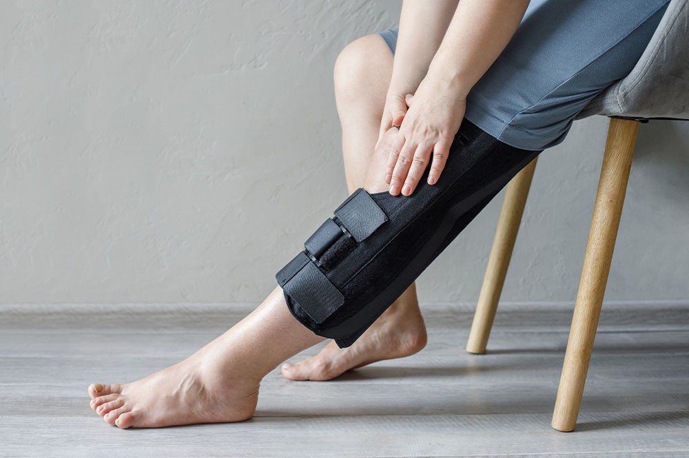
- braces
- functional wraps
- kinesio taping
- instrumental physiotherapy
- low-intensity diathermy
- pulsating magnetic field magnetotherapy
- high-energy lasers.
- a recovery of mobility
- increase in muscle tone
- restoration of proper movement kinematics (walking, running etc.)
- computerized proprioception (proboard)
- recovery of walking and/or athletic-sporting “gesture”
The rehabilitation program varies and, especially in athletes, must be managed by qualified personnel specialized in these types of pathologies who can change their approach at any time to optimize and adjust the rehabilitation program for optimal healing.
Conservative treatment is also a good strategy (pre-surgical treatment) in case of the likelihood of subsequent surgical treatment.
The individual with toned muscles, activated proprioception, and a deflated and uninflamed joint will face surgery in a certainly greatly improved condition, and their subsequent rehabilitation will be more effective and faster.
As soon as the load on the knee joint can be increased, physiotherapists integrate active mobilization exercises and elaborate with an individual training program.
Through a progressive accumulation of functional strengthening exercises and sensorimotor training, the motor function of the affected extremity is restored. One must also work with sensorimotor and proprioceptive training to establish a safe and biomechanically optimal gait.
The training program must continually adapt to the patient’s resilience and needs throughout therapy.
The prevention
The best prevention for not developing post-traumatic knee osteoarthritis would be never having a knee injury.
But aside from the obvious, there are some golden rules to keep a knee, whether healthy or previously damaged, at its best.
Because the knee joint is constantly exposed to stress in everyday life, it is imperative to take preventive measures and move gently for the joint. This relates to practicing knee-friendly behavior with simple actions and rules, supplemented with an exercise program that helps train specific muscle groups, thus protecting the knee joint through muscle strength and relieving it from load.
- Movement
- Decrease body weight according to BMI
- Relieve the knee joint
- Do not carry heavy loads
- Avoid sitting or standing for long periods
- Wear flat-heeled shoes that are comfortable and appropriate for the type of support
- Do only knee-friendly sports
- Train your leg muscles every day
In conclusion, it can be summed up as: “move a lot, exert little effort!”


